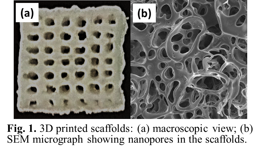

Tissue engineering and 3D-printing techniques it is possible to design a customized tracheal model with a morphology that is suitable for the patient and that supports the force to maintain the tissue engineered trachea (TET) shape (Sing et al., 2017). With advances in 3D-printing techniques, many studies have combined the two in order to improve the technologies that are available for tracheal surgeries. Tissue engineering is a promising approach to treat massive airway dysfunctions such as tracheomalacia or stenosis. Nevertheless, donor-site morbidity, erosion, infection, the requirement of immunosuppression in a cancer patient, as well as exceedingly complex laboratory and surgical techniques have prevented widespread clinical use (Fabre et al., 2013 Crowley et al., 2015 Friedman & Mayer, 1992 Propst et al., 2011). Several studies have turned to grafting technologies to overcome the clinical needs facing tracheal repair. However, performing such a procedure comes at a sacrifice of tracheal length for tracheal inner diameter.Īn innovative solution to overcoming these difficulties is the usage of artificial substitutes to replace long-segment narrowed trachea. Recently, slide tracheoplasty has improved outcomes significantly for inoperable patients (Wang et al., 2016 Basta et al., 2015). Tracheal resection and re-anastomosis have existed as a solution since late nineteenth century, but it was contraindicated for stenotic segments longer than 5 cm in adults and 2 cm in children for the risk of excess tension (Ho & Koltai, 2008). In mild-to-moderate tracheomalacia, observation or continuous positive airway pressure therapy might be effective yet, if these fail and the malacia is severe, surgery is frequently indicated (Carden et al., 2005). Both conditions are life-threatening for the patients (Chan et al., 2019 Wain Jr., 2009). Adult malacia and stenosis are typically related with chronic obstructive pulmonary disease, and can occur secondarily from tracheostomy, tumors, infection, prolonged intubation, trauma, and from external compression by vascular structures (Chan et al., 2019). Congenital forms of tracheomalacia and tracheal stenosis are more commonly seen in premature infants and are associated with severe symptoms (Kugler & Stanzel, 2014). Tracheal stenosis is a rare condition where there is a narrowing of the trachea that causes breathing problems. Tracheomalacia is defined as a soft cartilaginous sustenance of the trachea which can lead to the collapse and narrowing of the airway lumen during expiration. Nevertheless, additional long-term in vivo studies need to be performed to assess the efficacy and safety of tissue-engineered tracheal grafts in terms of mechanical properties, behavior and, the possibility of further stenosis development. Several strategies have been used to overcome the difficulty of airway reconstruction such as scaffold materials, construct designs, cellular types, biologic components, hydrogels and animal models used in tracheal reconstruction. 3D-bioprinting have contributed to current preclinical and clinical efforts in airway reconstruction. There are still some surgical limitations for certain pathologies, however tissue engineering is a promising approach to treat massive airway dysfunctions. Both conditions are life-threatening when not managed properly. In adulthood, are typically related with chronic obstructive pulmonary disease, and can occur secondarily from tracheostomy, prolong intubation, trauma, infection and tumors. Congenital tracheomalacia and tracheal stenosis are commonly seen in premature infants.


 0 kommentar(er)
